Invited Speaker

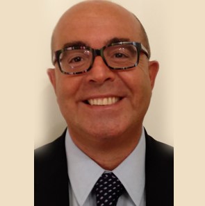
Dr. Andrea Tinelli
ProfessorDepartment of Obstetrics and Gynecology, “Veris delli Ponti” Hospital, Scorrano, Lecce, Italy;
Division of Experimental Endoscopic Surgery, Imaging,Technology and Minimally Invasive Therapy, Vito Fazzi Hospital, P.zza Muratore, Lecce, Italy;
Laboratory of Human Physiology,Phystech BioMed School, Faculty of Biological & Medical Physics, Moscow Institute of Physics and Technology (State University), Russia
Speech Title: Pseudocapsule Thickness in Reproductive Surgery: A Further Possible Correlation Between Submucous Fibroids And Fertility
Abstract: Uterine fibroid, during its growth, cause the progressive formation of a peripheral anatomical structure, a pseudocapsule. This structure originates fromthe fibroid compression of the surrounding myometrium and separates fibroid from the healthy myometrium. The pseudocapsule shifts the myometrial muscular fibers, maintaining the integrity and contractility of uterine musculature. Fibroid is structurally anchored to its pseudocapsule by connective bridges, but it lacks its own true vascular pedicle. Occasionally, bridges of collagen fibers and vessels anchoringfibroid to myometrium interrupting the pseudocapsule surface. Those physio pathological phenomena result in the formation of a clear cleavage plane either between myoma and the pseudocapsule, or between the pseudocapsule and the surrounding myometrium, as well. At the ultrastructural level, visualized by transmission electron microscopy (TEM), the pseudocapsule cells have the features of smooth muscle cells like the myometrium, indicating that the pseudocapsule is part of the myometrium compressed by the myoma. Moreover, pseudocapsule is plentiful of collagen fibers, neurofibers and blood vessels, as a neurovascular bundle surrounding fibroid. Genetically, the pseudocapsule of the myoma has the same biological structure as the myometrium and the biochemical growth factors evaluation showed intense angiogenesis in pseudocapsule vessels. Angiogenetic factorsidentified in the pseudocapsule vessels are already widely involved in the physiology of the myometrium and these substances are thought to have a pivotal role in wound healing and muscular innervation. Myometrial wound healing is an interactive, dynamic process involving neuromodulators, angiogenetic factors, neuropeptides, blood cells, extracellular matrix, and parenchymal cells that follows three complex and overlapping phases: inflammation, tissue formation, and tissue remodeling. In the physiology of these processes, they also fit also nervous system and its neurotransmitters, as Substance P (SP), Vasoactive Intestinal Peptide (VIP), neuropeptide Y (NPY), Oxytocin (OXT), Vasopressin (VP), PGP 9.5, calcitonin gene-related peptide (CGRP), growth hormone-releasing hormone (GHRH). They play a role in mediating inflammation and wound healing, involved in physiology and scar repair in different tissues, including uterine muscle. In regenerative processes associated to pseudocapsule sparing, neuropeptides and neurotransmitters are speculativelyinvolved also in wound healing. Moreover, growth factors present in the myoma pseudocapsule induce angiogenesis peripherally to myometrium. The intracapsular myomectomycan be done bylaparotomic, laparoscopic, robotic, vaginal and hysteroscopicapproach.The surgical benefit is visible during and after myomectomy: the bleeding is reduced, the myometrial anatomyis largely respected, themyometrial healing is preserved and enhanced, as confirmed by clinical and ultrasound investigations on scar site after intracapsular myomectomy.The study of the thickness of the pseudocapsule showed an increase in submucosal fibroids, compared to intramural and subserosal ones. This feature has a further impact on female reproduction to be investigated, as submucosal fibroids have been shown to negatively impact fertility.
Biography: Dr. Andrea Tinelli completed his medical degree magna “cum laude” in 1996, at School of Medicine “Federico II” of Naples, Italy, and specialized in Obsteric & Gynecology “cum laude” in 2000 at University of Trieste, Italy. He carried out surgical fellowship in University of Ljubljana (Slovenia), in Abteilung für Gynäkologie und Geburtshilfe, LKH Villach (Austria), in European Institute of Oncology (Milan, Italy) and in National Oncological Institute of Aviano (Italy).
Dr Tinelli currently is Chief of Obstetric & Gynecology Department at “Veris delli Ponti” Hospital, Scorrano, Italy and he’s Head of Division of Experimental Researches on Endoscopic Surgery, Imaging, Minimally Invasive Technology and Therapy, at “Vito Fazzi” Hospital, Lecce, Italy.
He’s a PhD in Morphological Molecular Sciences, at Catholic University of Sacred Heart, Rome, Italy. Dr. Tinelli has National Authorization as Full Professor in Obstetrics and Gynecology in Italy.
Dr Tinelli is Adjunct Professor at Moscow Institute of Physics and Technology (MIPT), Moscow, Russia and Honorary Professor at Jiaotong University, Xi'an, China.
He is permanent Board Member of D.R.e.A.M. Committee, at University of Salento, Italy.
Dr. Tinelli is author and co-author of 10 Books, more than 400 papers, on peer-reviewed medical journals and 70 textbook chapters; he collaborates with many Hospitals and Universities, as Editorial Board Member of Scientific Journals. The h-Index is 30, with more than 3700 citations.
His clinical specialties include minimally invasive surgery and gynecological endoscopy, obstetric surgery and complications management, gynecological surgical oncology, molecular biology in gynecology, endometriosis and pelvic floor repair.
